Age Related Macular Degeneration Part 1 – How To Detect

Source: Internet
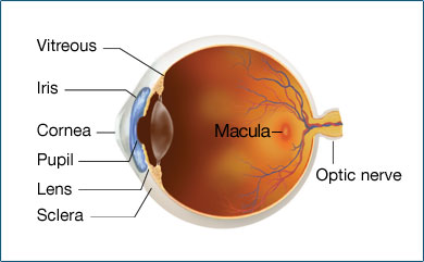
The Macula
The macula provides sharp, central vision and is made up of millions of light-sensing cells. It is one of the most sensitive part of the retina, which is located at the back of the eye. When the macula is damaged, the central vision tends to appear blurry, distorted, or dark.

Source: Internet
Age related macular degeneration: The Various Tests
AMD or age related macular degeneration usually start without symptoms. Only a comprehensive dilated eye exam can detect age-related macular degeneration. The eye exam shall include the following:
- Dilated eye exam. Generally drops are placed in the eyes to widen or dilate the pupils which provides a better view of the back of your eye. Using a special magnifying lens, he or she then looks at the retina for signs of AMD and other eye problems.
- Visual acuity test. To measure how well you see at distances an eye chart is used.
- Amsler grid. The eye care professional also may ask to look at an Amsler grid (The Amsler Grid looks like graph paper, with dark lines forming a square grid. Some versions have white lines on a dark background.) Changes in the central vision may also cause the lines in the grid to disappear or appear wavy.
- Fluorescein angiogram. Performed by an ophthalmologist, a fluorescent dye is injected into the arm and pictures are taken as the dye passes through the blood vessels in your eye. This makes it possible to see leaking blood vessels, which occur in a severe, rapidly progressive type of AMD. Sometimes complications to the injection can arise which is rare, and lead to nausea and severe allergic reactions.
- Optical coherence tomography. Just like ultrasound, which uses sound waves to capture images of living tissues. Optical Coherence Tomography is also similar except that it uses light waves, and provide high-resolution images of any tissues which are penetrated by light such as the eyes. The light beam is generally painless. After the eyes are dilated, you’ll be asked to place your head on a chin rest and hold still for several seconds while the Professional obtains the required images.
While the first signs of age related macular degeneration is almost nil, you should definitely get your eyes checked up frequently so that you can avail the required treatment in time. We will share more about age related macular degeneration in our upcoming blog.


